Leg Muscle Diagram Posterior : Muscles Of The Leg Part 2 Anterior And Lateral Compartments Anatomy Tutorial Youtube
Leg Muscle Diagram Posterior : Muscles Of The Leg Part 2 Anterior And Lateral Compartments Anatomy Tutorial Youtube. Deltoid triceps brachii brachioradialis extensor carpi ulnaris extensor carpi digitorum. This muscle diagram is interactive: Muscle anatomy leg muscles diagram muscle diagram nerves in leg. The muscle of the anterior compartment (arm in anatomical position) function as flexors while the muscles of the posterior compartment function as extensors. You'll learn about the muscles, bones, and other structures of each area of the leg.
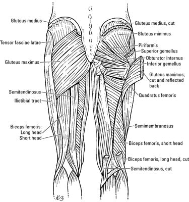
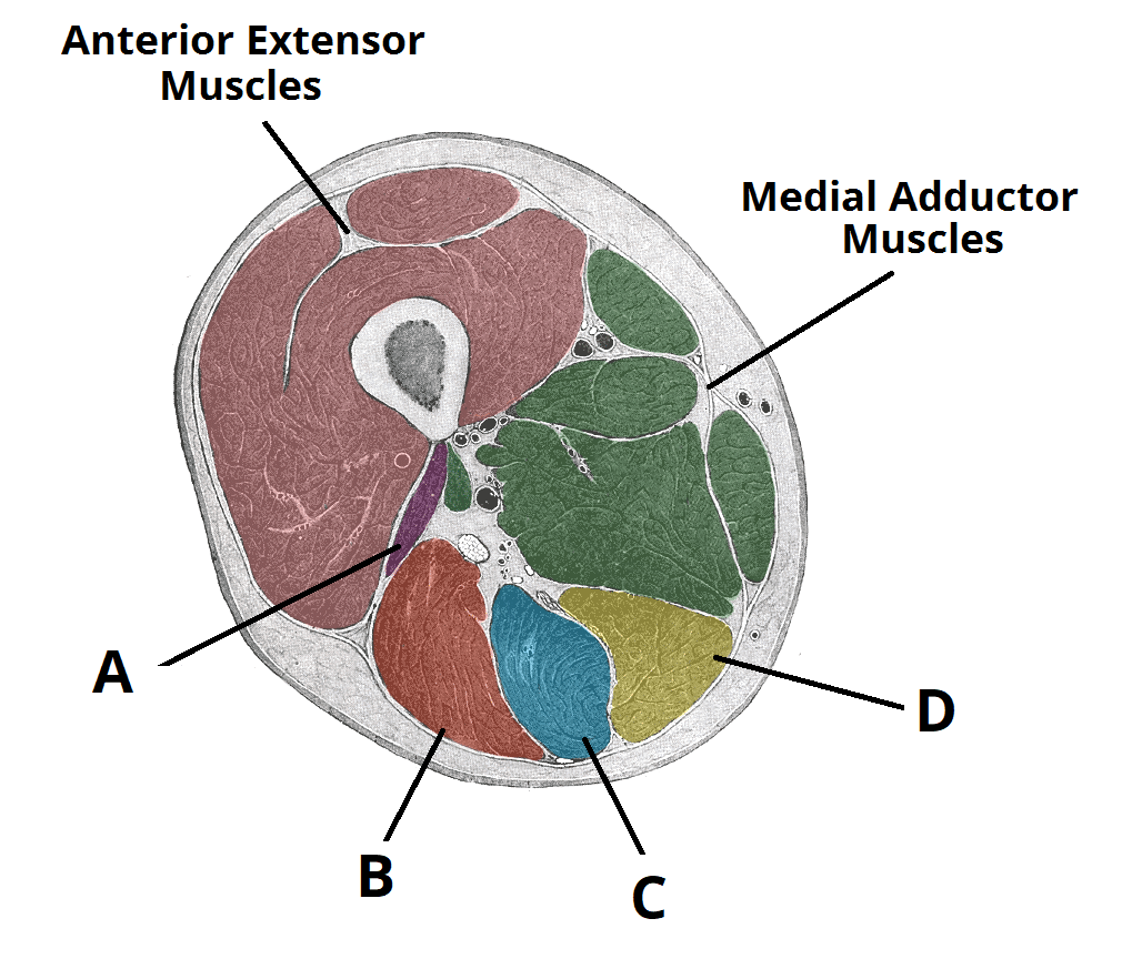
Rotator cuff muscle with anatomical posterior and anterior view expample. Get in touch with us today! This is the largest of the three compartments of the thigh. Leg muscle anatomy posterior leg muscles diagram photo album 10 / 10 ( 2 votes ) in this image, you will find tensor fascia latae, rectus femoris, vastus lateralis, iliopsoas, pectineus, adductor longus, gracilis, sartorius, vastus medialis, gluteus maximus, adductor magnus, semitendinosus, gracilis. The flexor hallucis longus muscle (fhl) is one of the three deep muscles of the posterior compartment of the leg that distally attaches to the plantar surface of the hallux (great or big toe).
You'll learn about the muscles, bones, and other structures of each area of the leg.
The large muscle of the posterior part of the lower leg. The flexor hallucis longus muscle (fhl) is one of the three deep muscles of the posterior compartment of the leg that distally attaches to the plantar surface of the hallux (great or big toe). Its action causes plantar flexion and inversion of. It contains the plantar flexors: Distinguishing feature soleus & obturator externus: The following diagram illustrates the actions of the terms adduction, abduction, flexion anterior compartment thigh muscles. Leg muscle anatomy posterior leg muscles diagram photo album 10 / 10 ( 2 votes ) in this image, you will find tensor fascia latae, rectus femoris, vastus lateralis, iliopsoas, pectineus, adductor longus, gracilis, sartorius, vastus medialis, gluteus maximus, adductor magnus, semitendinosus, gracilis. Anatomy muscle 3d illustration 3d rendering adductor magnus anatomical arthritis back biceps femoris body buttocks calf muscle diagram female fitness gastrocnemius glutes gluteus maximus gracilis. Muscles that move the leg. Deltoid triceps brachii brachioradialis extensor carpi ulnaris extensor carpi digitorum. Muscles of medial compartment of thigh. 3d medical illustration and rendering on leg posterior muscles for our client in australia. Tibialis posterior originates on the proximal 2/3 of tibia and fibula and inserts onto the medial cuneiform and navicular.
The muscle forms the floor of the popliteal fossa the muscle fibers extend through the posterior compartment of the leg and converge to form a solid tendon that passes behind the distal end of the. By dawn lewis the posterior sling is comprised of the latissimus dorsi muscle on one side of the body, the ipsilateral or same side thoracolumbar fascia and then. We'll break down the anatomy and function of the upper leg, knee, lower leg, ankle, and foot. Deltoid triceps brachii brachioradialis extensor carpi ulnaris extensor carpi digitorum. A muscle along the outside of the leg that bends the foot out at the ankle.

Superficial posterior leg compartment | origins, insertions, etc.
They allow you to move and provide support for your upper body. Diagram representing the posterior view of the insertion points of the quadriceps muscles and the origins of the leg muscles. The posterior compartment of the leg is one of the fascial compartments of the leg and is divided further into deep and superficial compartments. Here we explain the major muscles of the human body. Tibialis posterior originates on the proximal 2/3 of tibia and fibula and inserts onto the medial cuneiform and navicular. The following life study male figure sitting on the floor, shows a male the following life study lower torso and legs in a frontal view, shows the lower torso of a male figure. Distinguishing feature soleus & obturator externus: A muscle of the hip originating on the lateral surface of the ileum and inserted in the greater trochanter of the femur. Female hip and leg muscles labeled posterior view, 3d rendering. Each of these muscles is a discrete organ constructed of skeletal muscle tissue, blood vessels, tendons, and nerves. Your legs are two of your most important body parts. Muscles that move the leg. Posterior view of human muscular system.
A complete list of muscular system quizzes; Medially rotates leg when flexed. The flexor hallucis longus muscle (fhl) is one of the three deep muscles of the posterior compartment of the leg that distally attaches to the plantar surface of the hallux (great or big toe). The following life study male figure sitting on the floor, shows a male the following life study lower torso and legs in a frontal view, shows the lower torso of a male figure. The muscle of the anterior compartment (arm in anatomical position) function as flexors while the muscles of the posterior compartment function as extensors.
The muscle forms the floor of the popliteal fossa the muscle fibers extend through the posterior compartment of the leg and converge to form a solid tendon that passes behind the distal end of the.
Female hip and leg muscles labeled posterior view, 3d rendering. The large muscle of the posterior part of the lower leg. We'll break down the anatomy and function of the upper leg, knee, lower leg, ankle, and foot. 3d medical illustration and rendering on leg posterior muscles for our client in australia. They all insert into the calcaneus of the foot (the heel bone), via the. Here we explain the major muscles of the human body. Superficial posterior leg compartment | origins, insertions, etc. The following life study male figure sitting on the floor, shows a male the following life study lower torso and legs in a frontal view, shows the lower torso of a male figure. Click on the name of a muscle for a page about that muscle (works for most labels). Left leg, lateral (left) and posterior (right) views. Your legs are two of your most important body parts. Leg muscle anatomy posterior leg muscles diagram photo album 10 / 10 ( 2 votes ) in this image, you will find tensor fascia latae, rectus femoris, vastus lateralis, iliopsoas, pectineus, adductor longus, gracilis, sartorius, vastus medialis, gluteus maximus, adductor magnus, semitendinosus, gracilis. A muscle along the outside of the leg that bends the foot out at the ankle.

Post a Comment for "Leg Muscle Diagram Posterior : Muscles Of The Leg Part 2 Anterior And Lateral Compartments Anatomy Tutorial Youtube"